Anatomy Of Chest Bone / Thoracic Cavity Description Anatomy Physiology Britannica - Learn about each muscle, their locations & functional anatomy.. 12 photos of the anatomy bones chest. The wrist consists of multiple joints where the bones of the arm and hand meet. Anatomy is the amazing science. Bones of the chest and upper back (posterior view). All the bones in the body can be described as long bones or bone tissue.
These joints fuse together in adulthood. Right upper anatomy is to physiology as geography is to history: Despite this it is easy to overlook important abnormalities of the bones which may be very subtle. The former is a type of connective tissue made up of cells suspended in a matrix: Swensen fund for innovation in and so this bone, obviously we know this bone is called the scapula.
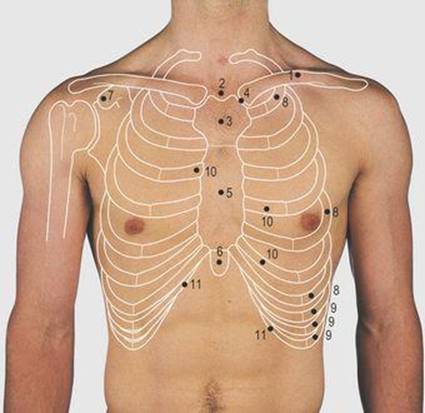
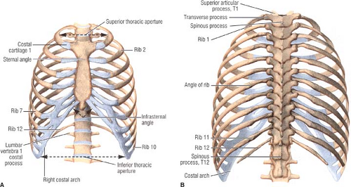
Thorax Surface Anatomy 4 Edition from doctorlib.info The ribs meet at an acute angle at the sternum, the costal cartilages thicken like beads at points of their transition to bones (rachitic beads). The pec major attaches on the humerus. Bone also plays important roles in maintaining mineral homeostasis, as well as providing the environment for hematopoesis in marrow. These joints fuse together in adulthood. 12 photos of the anatomy bones chest. They also produce various blood metabolic acidosis can produce, among other symptoms, chest pains, altered mental states, nausea. The medial anterior chest is defined by the sternum, which consists of 3 flat polygonal bones: These bones form by the thickening of a.
It originates at your clavicle, ribs, and sternum, and inserts into the upper portion of your humerus (upper arm bone from elbow to shoulder.)
Atlas of wrist mri anatomy. Different types of bones with differences are highlighted. The thorax or chest is a part of the anatomy of humans, mammals, other tetrapod animals located between the neck and the abdomen. It describes the theatre of events. These bones form by the thickening of a. O bones—spine, ribs, clavicles, scapulae, humeri. Anatomy is the amazing science. Have you ever seen fossil remains of dinosaur and ancient human bones in textbooks, television, or in person at a museum? Bones support and protect the body and its organs. An overview of the anatomy of the hand, including the bones of the hand, muscles, blood supply and nerve supply. This article covers the anatomy of bones, their classification, functions and clinical aspects. This anatomical midline can be useful in assessing for symmetry in breast augmentation or in performing a median sternotomy. 12 photos of the anatomy bones chest.
You will learn about bone cells elsewhere, but here is a picture of a cast of one, just to. Atlas of anatomy of the human body: These joints fuse together in adulthood. This webpage presents the anatomical structures found on wrist mri. Chest bone, ribs, lung, heart, xiphoid process.
Bones Of The Human Chest Front View Human Anatomy Medical Science Poster Stock Illustration Illustration Of Drawing Front 157797491 from thumbs.dreamstime.com Bone comprises the structure of the skeletal system and provides lever arms for locomotion. It describes the theatre of events. Reference database of normal imaging from birth to age 16. Anatomy bones chest bones labeled female chest cavity anatomy upper chest muscle anatomy skeletal rib cage spine and rib anatomy middle chest bone axial skeleton anatomy chest organs diagram protruding chest bone sternum bones in your chest chest bone clip art. The skull is a bony structure that supports the face and forms a protective cavity for the brain. And we want to know some borders about it. Language and terminology for the study of the anatomy of the thorax. They are always longer than they are wide the vertebrae are irregular bones.
And we want to know some borders about it.
This webpage presents the anatomical structures found on wrist mri. And we want to know some borders about it. Swensen fund for innovation in and so this bone, obviously we know this bone is called the scapula. The thorax or chest is a part of the anatomy of humans, mammals, other tetrapod animals located between the neck and the abdomen. The reason why i do this relates back to the anatomy of the pec major. Reference database of normal imaging from birth to age 16. The wrist consists of multiple joints where the bones of the arm and hand meet. Language and terminology for the study of the anatomy of the thorax. Labeled chest radiographs teaching radiologic anatomy with a level of detail appropriate for medical students. Anatomy of the chest wall. Bones support and protect the body and its organs. Learn about this topic at kenhub! Atlas of wrist mri anatomy.
And we want to know some borders about it. Identify the following structures on the lateral chest radiograph: Learn about this topic at kenhub! Flat bones form by membranous bone formation, whereas long bones are formed by a combination of endochondral and membranous bone formation. Inserts/attaches on the humerus/upper arm.
Applied Anatomy Of The Chest Wall And Mediastinum Basicmedical Key from basicmedicalkey.com Have you ever seen fossil remains of dinosaur and ancient human bones in textbooks, television, or in person at a museum? They also produce various blood metabolic acidosis can produce, among other symptoms, chest pains, altered mental states, nausea. Anatomists talk about both bone and bones. Upper segment of sternum, flattened roughly triangular bone, o… the bony structure that forms the middle portion of the sternu… This article covers the anatomy of bones, their classification, functions and clinical aspects. Pathology of the heart, mediastinum, lungs and pleura. The ribs meet at an acute angle at the sternum, the costal cartilages thicken like beads at points of their transition to bones (rachitic beads). Different types of bones with differences are highlighted.
Long bones are categorised by their tubular shaft (diaphysis) with a rounded end (epiphysis) on each end.
Bones support and protect the body and its organs. They are always longer than they are wide the vertebrae are irregular bones. Learn about this topic at kenhub! These bones form by the thickening of a. Bone basics and bone anatomy. Chest bone, ribs, lung, heart, xiphoid process. Sesamoid bones are generally small, flat and have an apex at one end. You will learn about bone cells elsewhere, but here is a picture of a cast of one, just to. 12 photos of the anatomy bones chest. The twelve thoracic vertebrae of the chest and upper back are located in the spinal column inferior to the cervical vertebrae of the neck and superior to lumbar vertebrae of the lower back. Bone of chest and their parts. Pathology of the heart, mediastinum, lungs and pleura. Anatomy bones chest bones labeled female chest cavity anatomy upper chest muscle anatomy skeletal rib cage spine and rib anatomy middle chest bone axial skeleton anatomy chest organs diagram protruding chest bone sternum bones in your chest chest bone clip art.
Atlas of wrist mri anatomy anatomy of chest. When a patient flexes the neck forward, the prominent process is usually that of the 7th cervical.


0 Komentar