Muscles Anterior Full Body Diagram - Muscles Anterior Full Body Diagram Anterior Muscle Diagram Untpikapps - Click on the name of a muscle for a page about that muscle (works for most labels).
Muscles Anterior Full Body Diagram - Muscles Anterior Full Body Diagram Anterior Muscle Diagram Untpikapps - Click on the name of a muscle for a page about that muscle (works for most labels).. This muscle diagram is interactive: Muscle anatomy chest 12 photos of the muscle anatomy chest anterior chest muscle anatomy, chest muscle anatomy and exercises, chest muscle anatomy male, chest wall muscle anatomy mri, female chest muscle anatomy diagram. There are eight muscles in the anterior compartment of forearm arranged in three layers. When you are taking anatomy and physiology you will be required to identify major muscles in the human body. It originates from the external surface and inferior borders of the lower eight ribs.
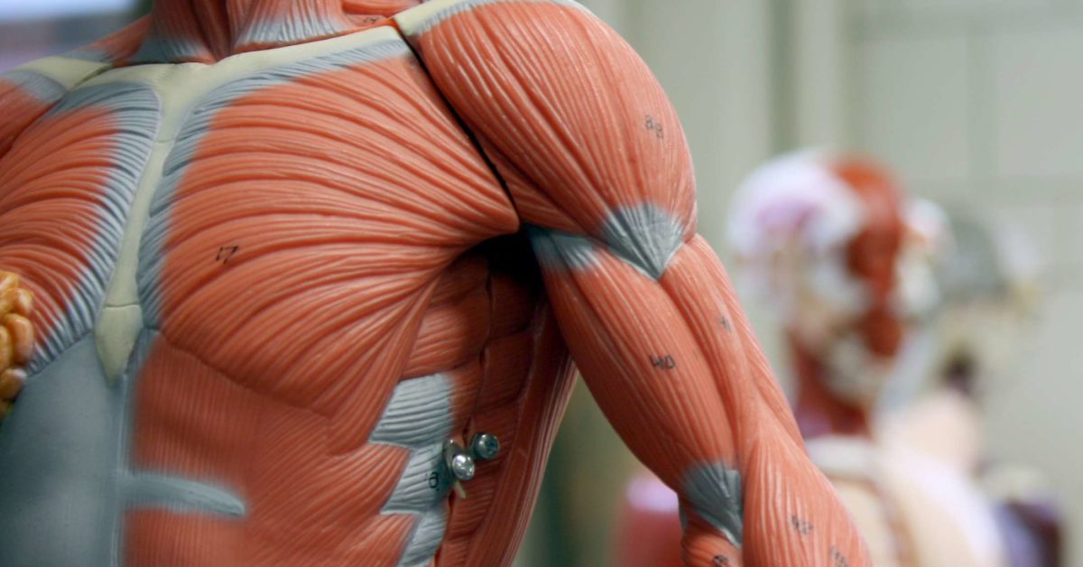
Serratus anterior, with deltoid muscle. This section explores the different types of muscles in our body and their involvement in sporting activities. Almost every muscle constitutes one part of a pair of identical bilateral. The muscles labelled in the anterior muscles diagram shown above are listed in bold in the following table Pectoralis major, pectoralis minor, serratus anterior, subclavius, external intercostals, internal intercostals, innermost intercostals the anterior trunk muscles cover the anterolateral part of the trunk by attaching to the bony framework of the thoracic cage and pelvis.
Pin On What Is from i.pinimg.com Click on the name of a muscle for a page about that muscle (works for most labels). Muscle anatomy chest 12 photos of the muscle anatomy chest anterior chest muscle anatomy, chest muscle anatomy and exercises, chest muscle anatomy male, chest wall muscle anatomy mri, female chest muscle anatomy diagram. Left ventricle and papillary muscles. Interactive human muscular system full body. Learn faster with these free muscle labeling diagrams. This is a table of muscles of the human anatomy. There are around 650 skeletal muscles within the typical human body. The muscles labelled in the anterior muscles diagram shown above are listed in bold in the following table
Get in touch with us today!
Anterior muscles in the body. Skeletal muscles rarely work by themselves to achieve movements in the body. Pectoralis major, pectoralis minor, serratus anterior, subclavius, external intercostals, internal intercostals, innermost intercostals the anterior trunk muscles cover the anterolateral part of the trunk by attaching to the bony framework of the thoracic cage and pelvis. Superficial and deep anterior muscles of upper body. The sartorius is the longest muscle in the body. Superficial and deep anterior muscles of upper body. The muscular system consists of various types of muscle that each play a crucial role in the function of the body. More often they work in groups to produce precise movements. Arm anterior 3d illustration project. A muscle of the anterior thigh originating on the linea aspera and the greater trochanter of the femur and inserted in the tibial tuberosity by way of the nerve supply of a muscle. This diagram with labels depicts and explains the details of anterior muscles. Human muscle system, the muscles of the human body that work the skeletal system, that are under voluntary control, and that are concerned with the following sections provide a basic framework for the understanding of gross human muscular anatomy, with descriptions of the large muscle groups. Serratus anterior, with deltoid muscle.
Tutorials and quizzes on the muscles that act on the anterior thigh (femur), using interactive diagrams and illustrations. Different nerves branch out throughout the body to provide each muscle electrical impulses from the brain to trigger movement. This muscle diagram is interactive: A muscle of the anterior thigh originating on the linea aspera and the greater trochanter of the femur and inserted in the tibial tuberosity by way of the nerve supply of a muscle. This section explores the different types of muscles in our body and their involvement in sporting activities.
Testosterone Anatomical And Biological Body Diagram With Brain Royalty Free Cliparts Vectors And Stock Illustration Image 102408474 from previews.123rf.com There are anterior muscles diagrams and posterior muscles diagrams. The muscles labelled in the anterior muscles diagram shown above are listed in bold in the following table Anterior muscles in the body. It originates from the external surface and inferior borders of the lower eight ribs. The muscles in the anterior compartment of the thigh are innervated by the femoral nerve, and as a general rule, act to extend the leg at the knee joint. This is a table of skeletal muscles of the human anatomy. Related posts of muscles in your body diagram. Muscles allow a person to move.
The sartorius is the longest muscle in the body.
On the next diagram we will indicate the intermediate layer of anterior compartment of forearm. This is a table of skeletal muscles of the human anatomy. Interactive human muscular system full body. This system is mainly concerned with producing movement through muscle contraction. This muscle diagram is interactive: Click on the name of a muscle for a page about that muscle (works for most labels). Arm anterior 3d illustration project. Superficial and deep anterior muscles of upper body. Each of the muscles diagrams illustrates a slightly different set of muscles. The sartorius is the longest muscle in the body. Identify the muscle labeled as 8 in the diagram above: Anatomy muscle man didactic abdominus transversalis achilles (calcaneal) tendon adductor brevis adductor longus adductor magnus biceps brachii biceps femoris brachioradialis coraco brachialis (under biceps. Produce wrist and/or finger flexion.
Start studying anterior muscles full body. Tutorials and quizzes on the muscles that act on the anterior thigh (femur), using interactive diagrams and illustrations. The muscles that affect the knee's movement run along the thigh and calf. The sartorius is the longest muscle in the body. The muscles labelled in the anterior muscles diagram shown above are listed in bold in the following table
11 Functions Of The Muscular System Diagrams Facts And Structure from post.medicalnewstoday.com These include mobility, stability, posture, circulation, digestion, and more. Arm anterior muscles labeled 3d illustration. The muscular system consists of various types of muscle that each play a crucial role in the function of the body. The muscular system consists of various types of muscle that each play a crucial role in the function of the body. Anterior and posterior muscles of the upper arm. Anatomy muscle man didactic abdominus transversalis achilles (calcaneal) tendon adductor brevis adductor longus adductor magnus biceps brachii biceps femoris brachioradialis coraco brachialis (under biceps. Skeletal muscles rarely work by themselves to achieve movements in the body. Muscles of the anterior compartment of the forearm.
Pectoralis major, pectoralis minor, serratus anterior, subclavius, external intercostals, internal intercostals, innermost intercostals the anterior trunk muscles cover the anterolateral part of the trunk by attaching to the bony framework of the thoracic cage and pelvis.
The muscular system consists of various types of muscle that each play a crucial role in the function of the body. Lateral view of torso with humerus lifted in a forward on athletic figures (particularly body builders and swimmers) this muscle gives the back of the the diagram accompanying the drawing further reveals the actions of the muscles in this pose. There are around 650 skeletal muscles within the typical human body. Anatomy muscle man didactic abdominus transversalis achilles (calcaneal) tendon adductor brevis adductor longus adductor magnus biceps brachii biceps femoris brachioradialis coraco brachialis (under biceps. This section explores the different types of muscles in our body and their involvement in sporting activities. Skeletal muscles rarely work by themselves to achieve movements in the body. Produce wrist and/or finger flexion. Anterior full body muscle diagram. Identify the muscle labeled as 8 in the diagram above: Muscle anatomy chest 12 photos of the muscle anatomy chest anterior chest muscle anatomy, chest muscle anatomy and exercises, chest muscle anatomy male, chest wall muscle anatomy mri, female chest muscle anatomy diagram. The muscles that affect the knee's movement run along the thigh and calf. Serratus anterior, with deltoid muscle. Muscle tissue is also found inside of the heart digestive organs.


0 Komentar